
Who dominates, and why
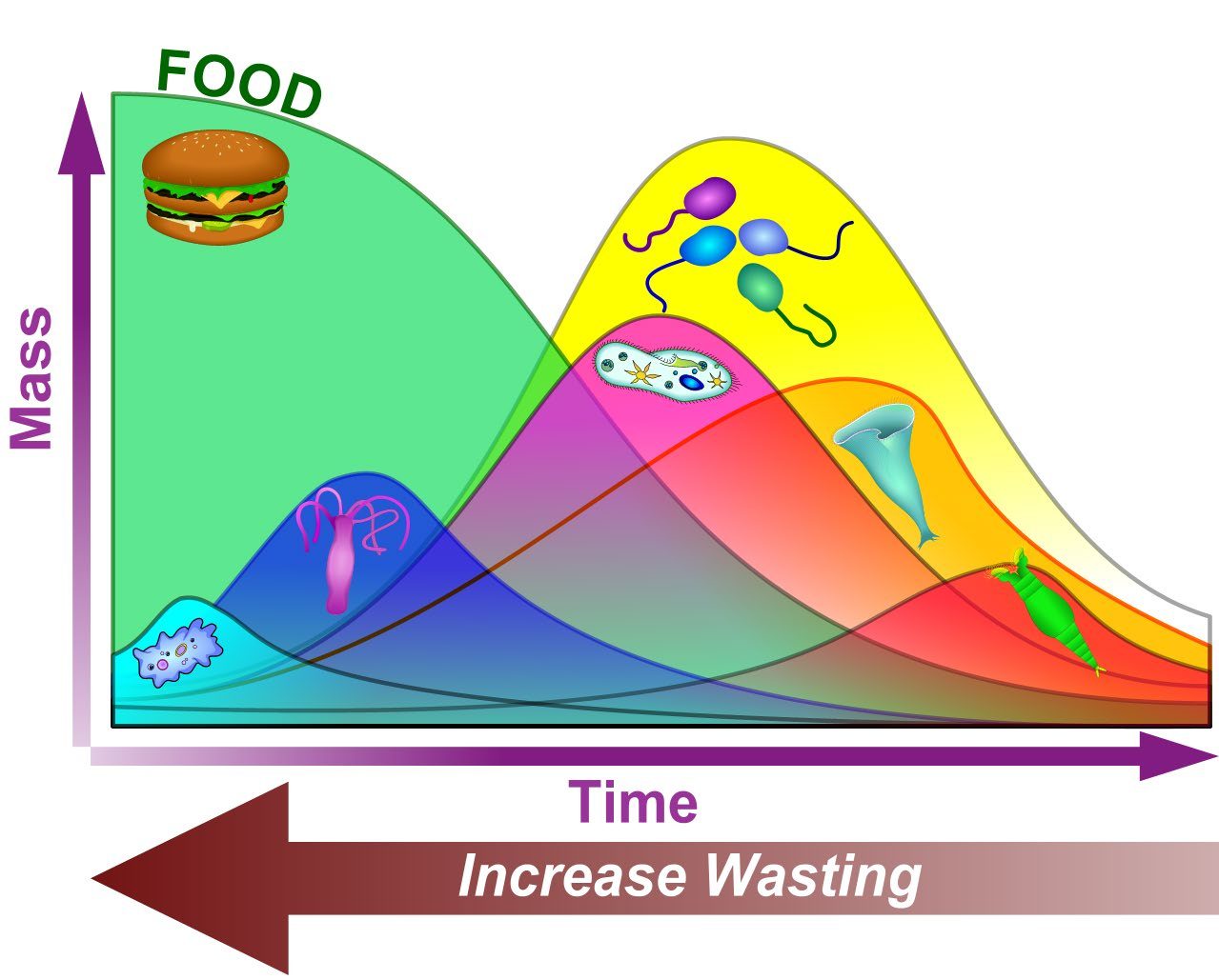
What you see, what it may indicate, and what process data to check next.
The microscope shows what the lab numbers cannot: floc structure, filaments and the higher life forms that reflect sludge age and process health.
Answer a few questions about what is on the slide and get pointed to the likely match.
Bell shaped ciliates attached to floc by a stalk. Vorticella, Epistylis, Opercularia, Carchesium.
Ciliates that swim between flocs eating dispersed bacteria. Paramecium, Litonotus, Aspidisca (crawler).
Multicellular animals with a crown of cilia and a forked foot. A sign of older, stable sludge.
Shape changing naked amoebae and shelled (testate) amoebae.
Small cells moving with one or more whip like flagella. Abundant in young sludge.
Small, thrashing roundworms. Usually long SRT and stable conditions.
Long chains of bacteria. A few support floc; too many cause bulking and foam.
Size, shape, strength and background clarity of activated sludge floc.
Bacteria growing as single cells or tiny clumps instead of floc. Cloudy effluent.
53 photomicrographs from Brad Musick's slide collection: protozoa, higher life forms, filaments, floc and stains.
10 original drawings: protozoa groups, higher life forms, floc structure, bacterial shapes, capsules and sheaths, respiration.