53 photomicrographs from Brad Musick's microscope slide collection, with what each one tells an operator. Select a photo to open it full size. Original drawings fill in what the photos do not show; see all of them on the illustrations page.
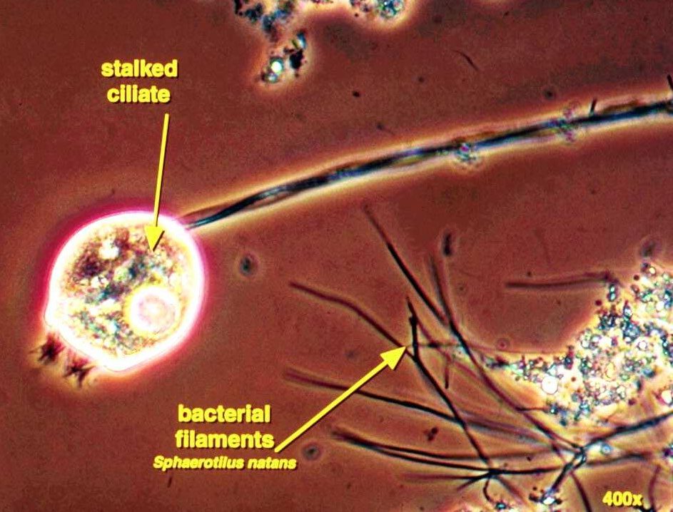
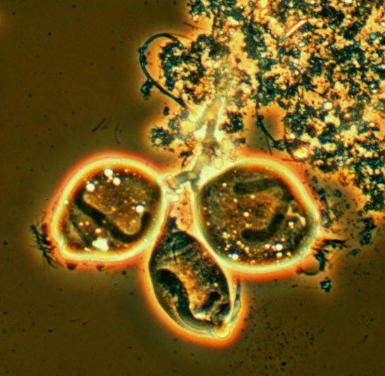
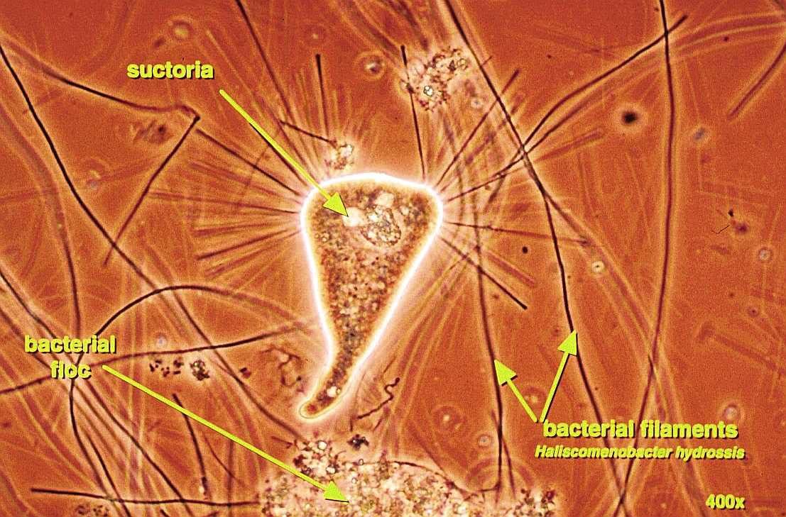
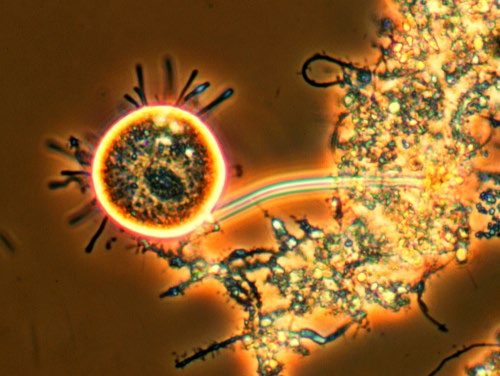
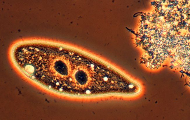
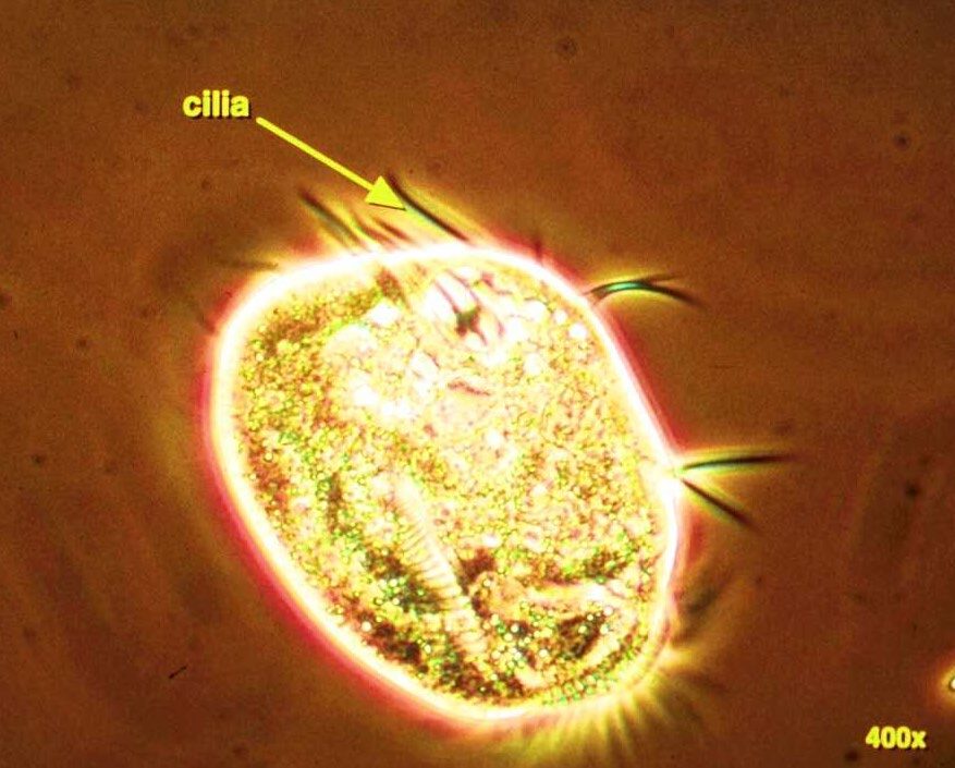
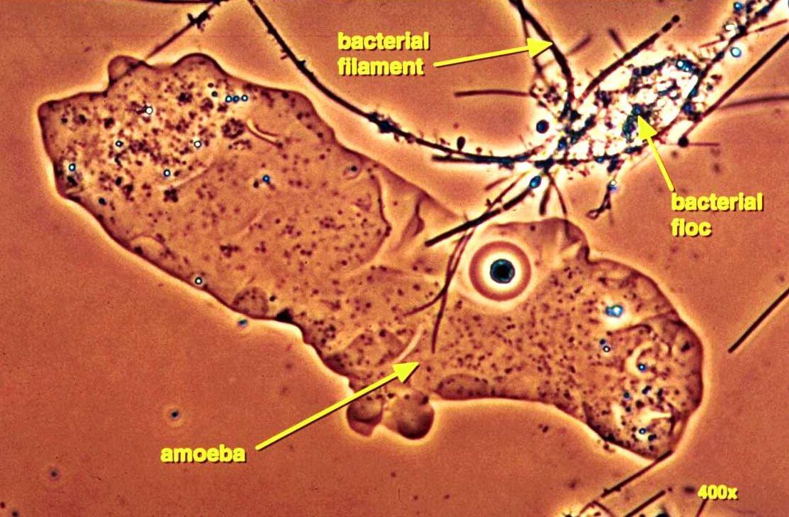
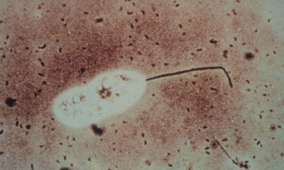
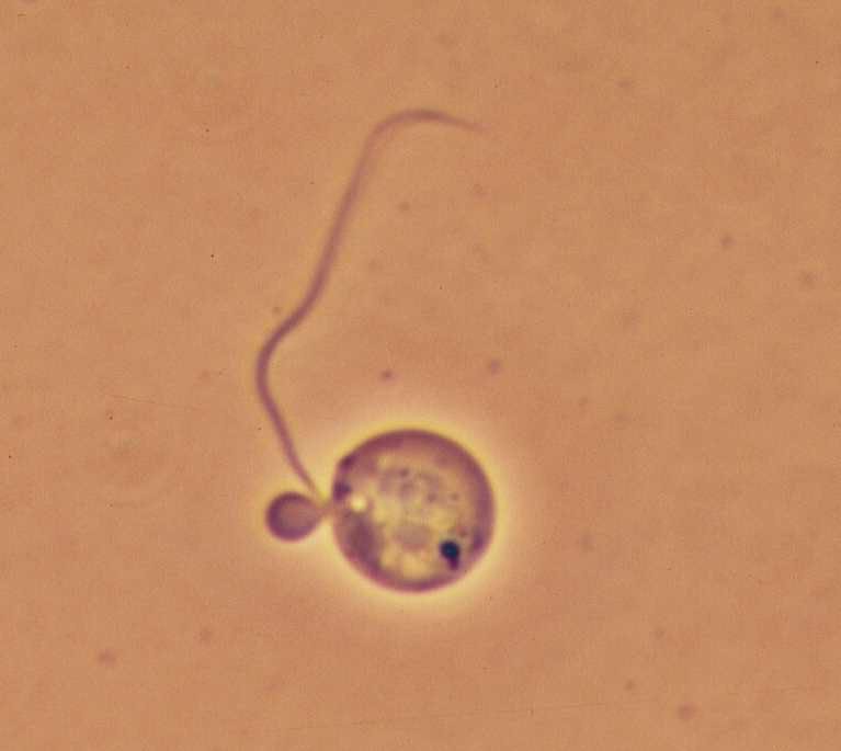
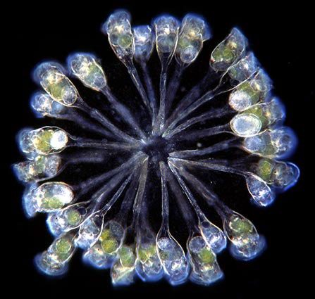
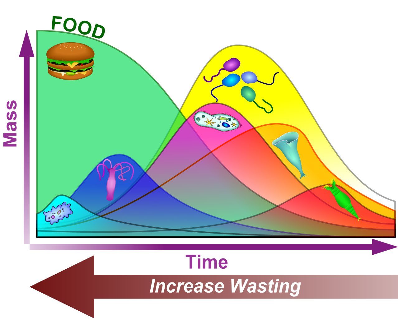
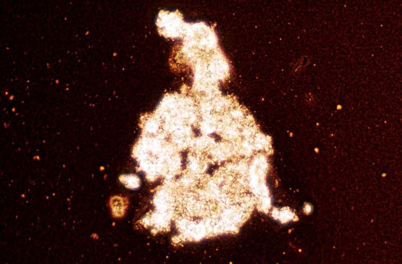
Stalked ciliate. Stalked ciliate attached by its stalk, with Sphaerotilus natans filaments in the same field. Phase contrast, 400x. What this indicates: Stalked ciliates point to a stable, mature sludge. S. natans in the same field is a low DO indicator, so check the DO profile.Stalked ciliates on floc. A group of stalked ciliates on the edge of a firm floc. Phase contrast. What this indicates: Healthy, open bells on firm floc usually go with good settling and a clear supernatant.Stalked ciliate, brightfield. Single stalked ciliate seen in brightfield. What this indicates: One healthy stalked ciliate on its own says little; count them across the slide and watch the trend.Suctorian. Suctorian (a stalked relative of the ciliates that catches prey with tentacles) among Haliscomenobacter hydrossis filaments. Phase contrast, 400x. What this indicates: Suctorians feed on other protozoa and usually show up with stalked ciliates in a stable, older sludge.Suctorian on floc. Round suctorian with tentacles on a floc. Phase contrast. What this indicates: Like stalked ciliates, suctorians are a sign of a mature, stable sludge.Free-swimming ciliate. Slipper shaped free-swimming ciliate beside a floc. Phase contrast. What this indicates: Free swimmers feed on dispersed bacteria. Many of them often means a younger sludge or a cloudy background.Crawling ciliate. Crawling ciliate with its cirri (fused cilia) visible. Phase contrast, 400x. What this indicates: Crawlers grazing on floc, together with stalked ciliates, suggest a stable, well flocculated sludge.Naked amoeba. Large naked amoeba beside a bacterial floc and filament. Phase contrast, 400x. What this indicates: Naked amoebae in numbers suggest a young sludge, a startup or recovery after an upset. One or two on a slide is normal.Flagellate. Flagellate with its long flagellum among dispersed bacteria. Brightfield. What this indicates: Flagellates with a cloudy, bacteria filled background point to young sludge or a high soluble BOD load.Small flagellate. Small round flagellate with one flagellum. Brightfield. What this indicates: Small numbers are normal. If flagellates dominate the slide, check F/M, SRT and any recent upset.Colonial stalked ciliates. A colony of stalked ciliates whose stalks branch from a single base, forming a rosette. Darkfield illumination. What this indicates: Large colonies of stalked ciliates take time to build, so they point to a stable, mature sludge with a steady load and good DO.Relative predominance of organisms. As food is used up over time (longer sludge age, lower F/M), the dominant organisms shift from amoebae (first curve) and flagellates (second curve) to free-swimming ciliates, then stalked ciliates and finally rotifers. Wasting more sludge pushes the population back toward the left. What this indicates: Which group dominates your slide tells you roughly where the sludge sits on this curve. Many flagellates and amoebae mean a young, high F/M sludge; stalked ciliates and rotifers mean an older, low F/M sludge.Graphic: Brad Musick
Illustrated
The six protozoa groups. Original line drawings of amoebae, flagellates, free-swimming, crawling and stalked ciliates and suctorians. What this indicates: Which group you see most of hints at sludge age: amoebae and flagellates for young sludge, crawling and stalked ciliates and suctorians for mature sludge.Original illustration for this site, not to scale.Naked and testate amoebae. A naked amoeba with pseudopods and vacuoles, and two shelled (testate) amoebae, Arcella and Euglypha. What this indicates: Many naked amoebae suggest a young sludge or a recovery from upset; testate amoebae suggest a long SRT, low F/M and often full nitrification.Original illustration for this site, not to scale.How small flagellates are. A 100x field with floc, and a 400x close up of one flagellate with its whip like flagellum. What this indicates: Flagellates are easy to miss at 100x. Large numbers at 400x point to high soluble BOD, a young sludge or low DO.Original illustration for this site, not to scale.
Rotifers and other higher life forms
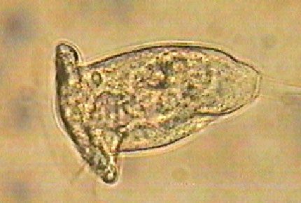
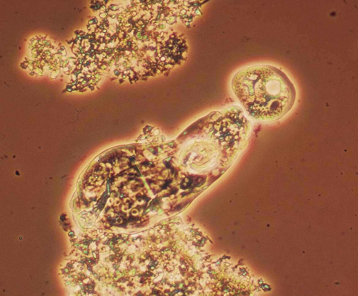
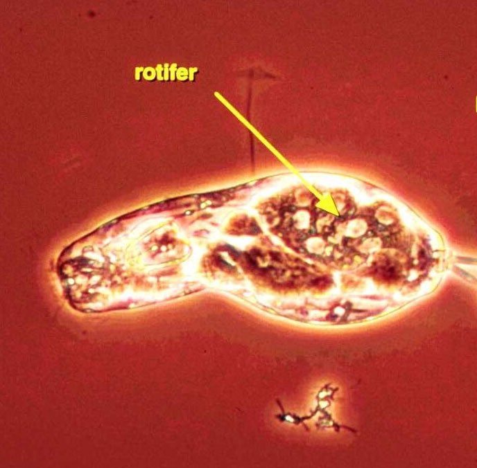
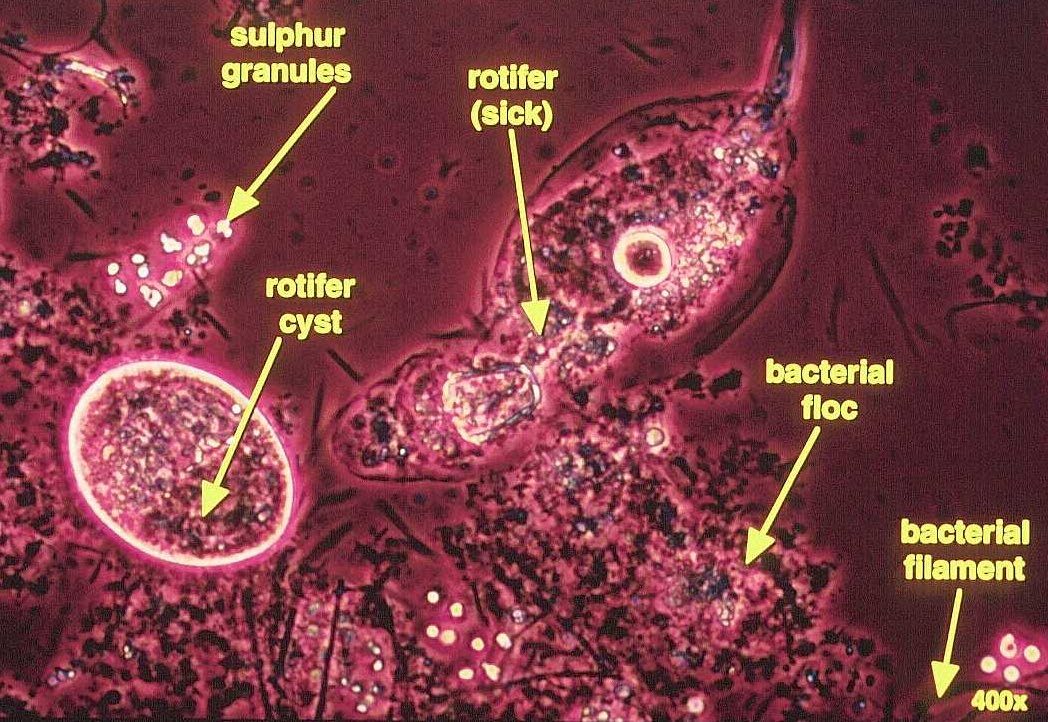
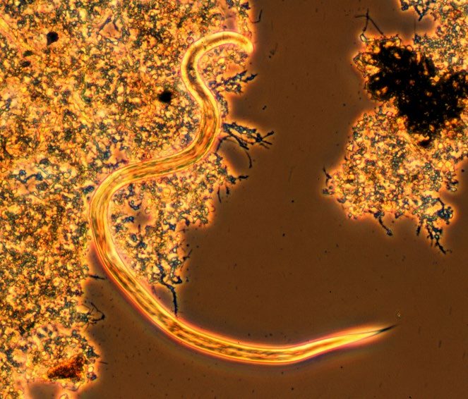
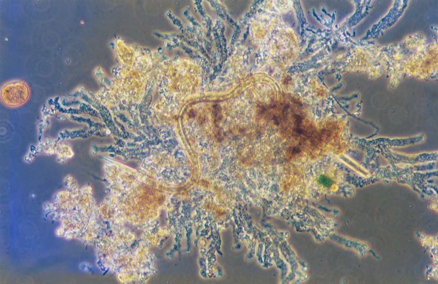
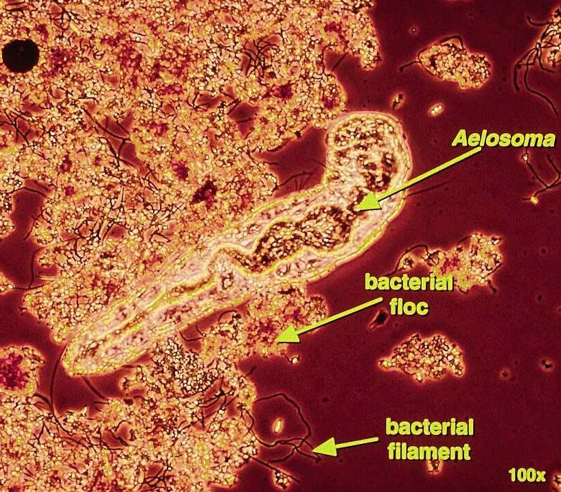
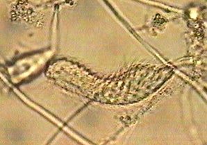
Rotifer on floc. Rotifer anchored to a floc by its foot. Phase contrast, 400x. What this indicates: Active rotifers point to a mature, stable sludge at moderate to long SRT.Rotifer. Rotifer showing its internal organs. Phase contrast. What this indicates: Rotifers need stable conditions. Large numbers with small, dense floc can mean the SRT is very long.Rotifer and cyst, sulfide toxicity. Sick rotifer and a rotifer cyst with sulfur granules nearby, from a plant with sulfide toxicity. Phase contrast, 400x. What this indicates: Rotifers forming cysts or losing activity, with sulfur granules on the slide, point to sulfide or another toxic stress. Check septicity and influent sulfide.Nematode. Nematode moving between flocs. Phase contrast. What this indicates: Nematodes are usually a sign of long SRT and stable conditions, common in extended aeration.Fingered Zoogloea and nematode. Fingered Zoogloea with a nematode in the same floc. Phase contrast. What this indicates: Fingered Zoogloea grows in high F/M systems where food is soluble or easily degraded and pH is low. Nematodes go with an older sludge.Bristle worm (Aeolosoma). Aeolosoma, a segmented bristle worm, among floc and filaments. Phase contrast, 100x. What this indicates: Bristle worms, like nematodes, are higher life forms that point to an older, stable sludge.Chaetonotus (gastrotrich). Chaetonotus, a ciliated gastrotrich. What this indicates: An uncommon higher life form, usually in older sludge.
Illustrated
Higher life forms. Rotifer, nematode, bristle worm (Aeolosoma) and water bear (tardigrade), with typical sizes. What this indicates: These multicellular animals grow slowly, so steady numbers usually mean a long SRT and a stable, well oxidized sludge.Original illustration for this site, not to scale.
Filamentous organisms
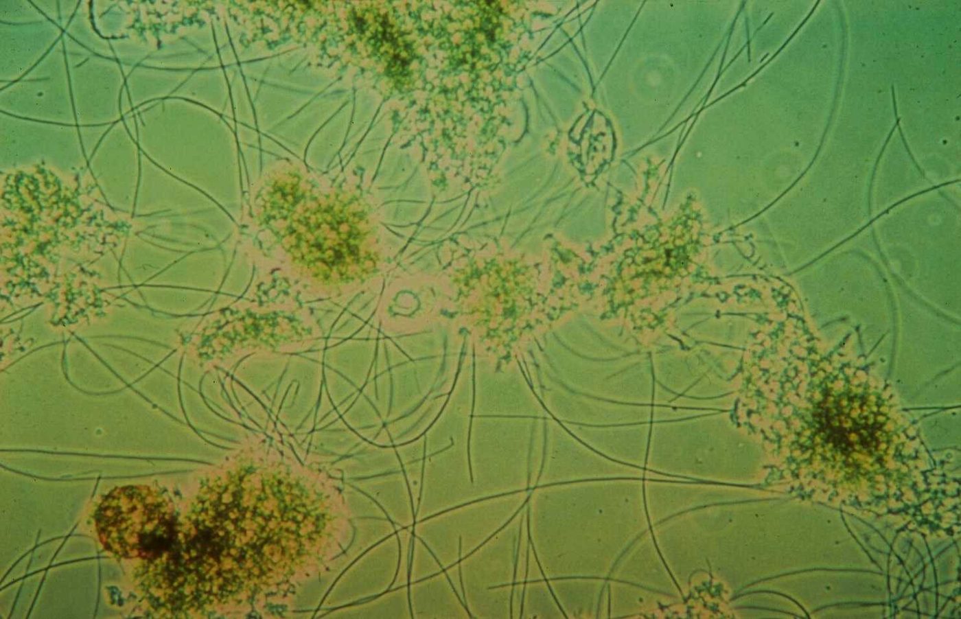
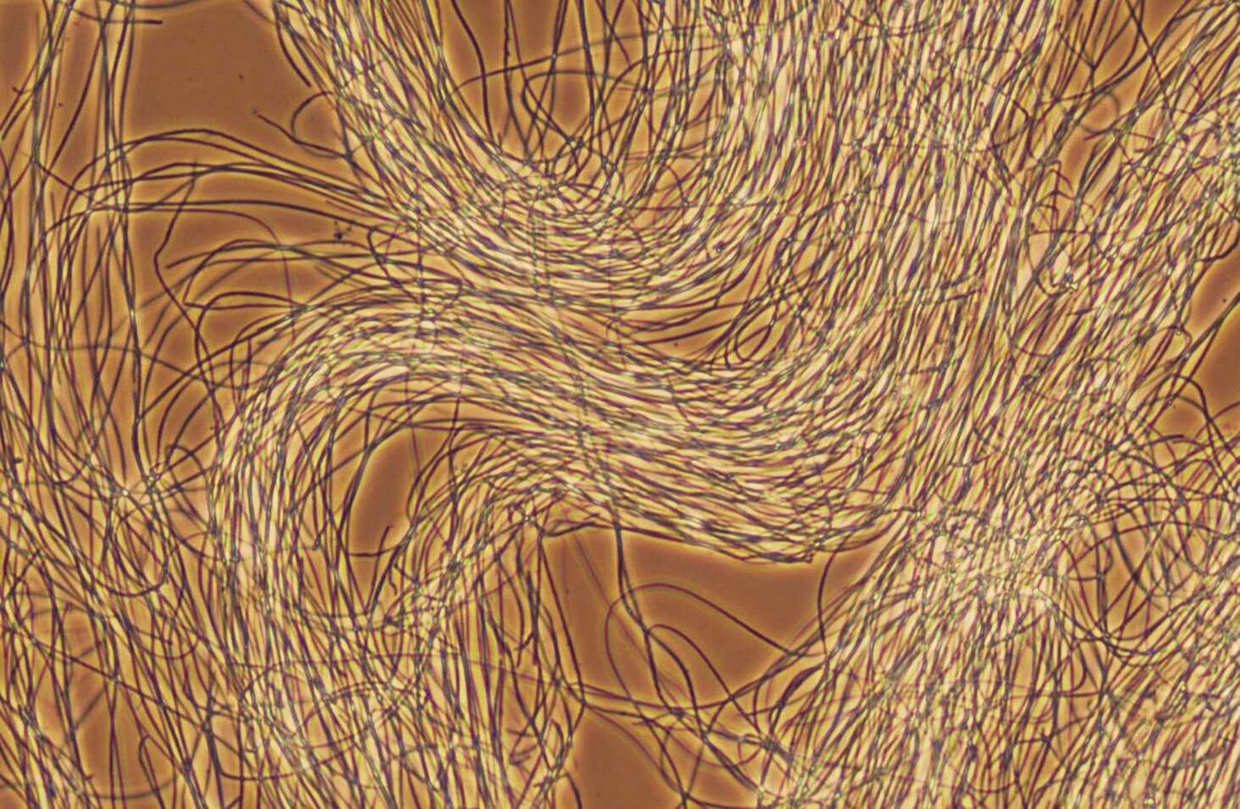
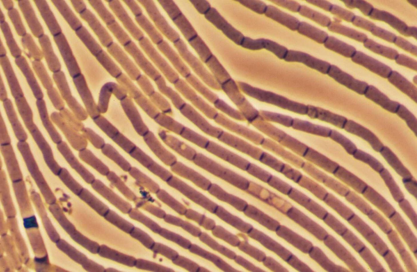
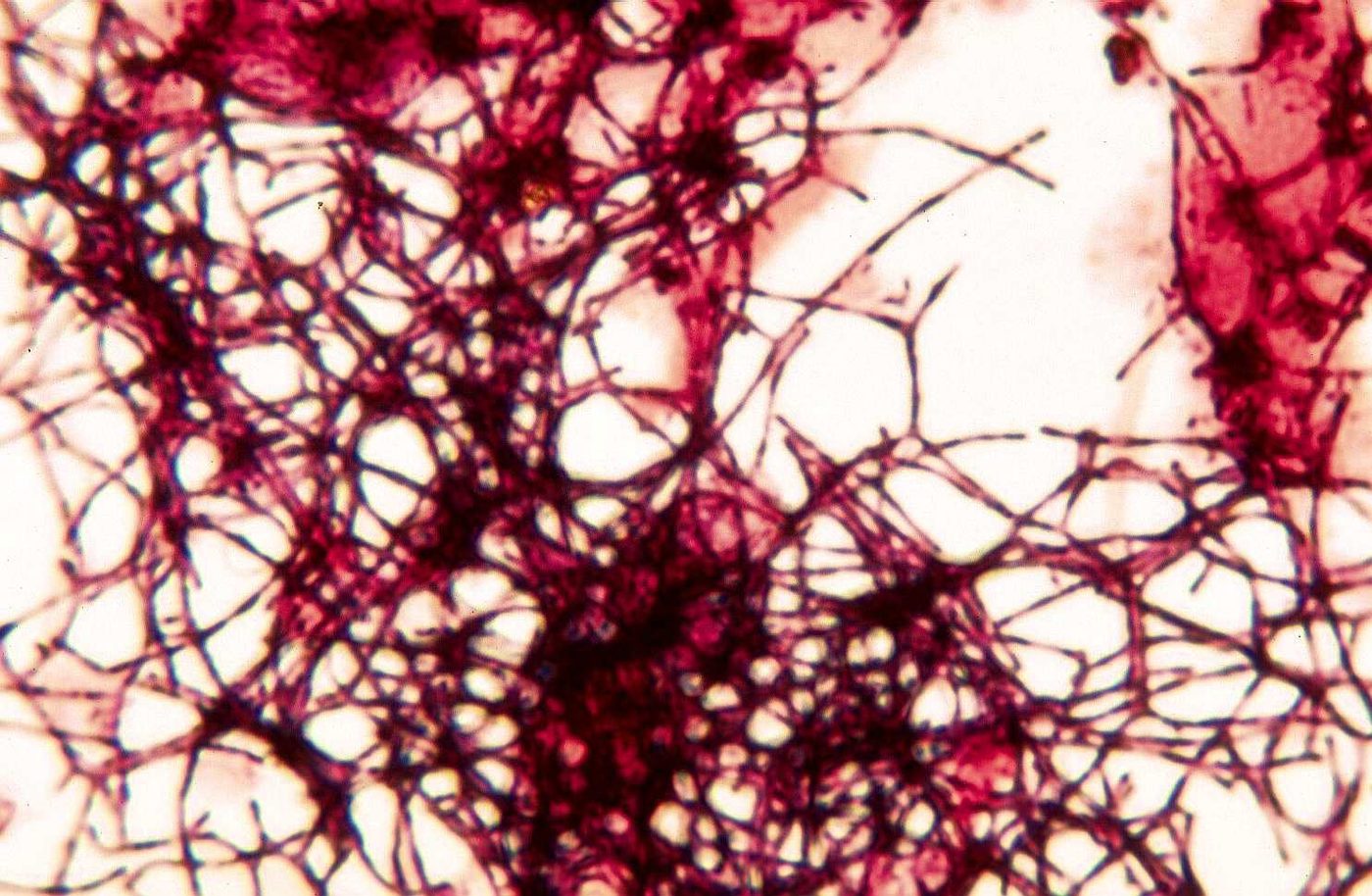
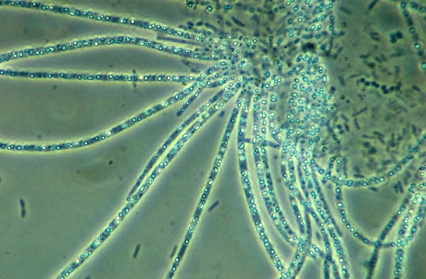
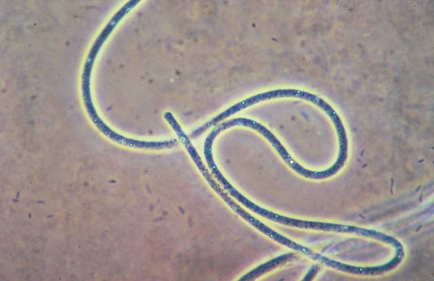
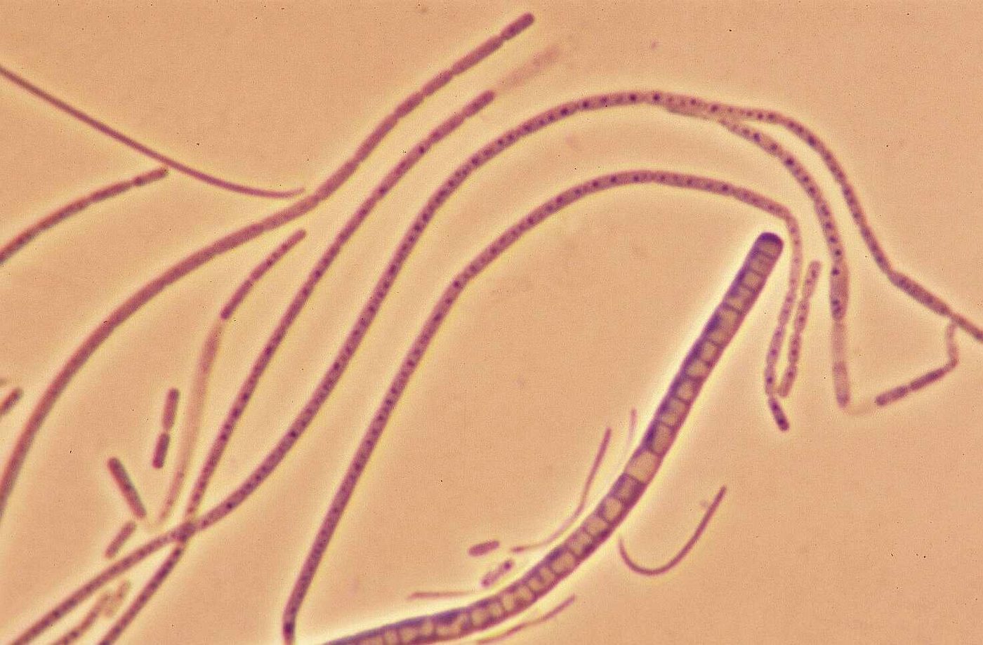
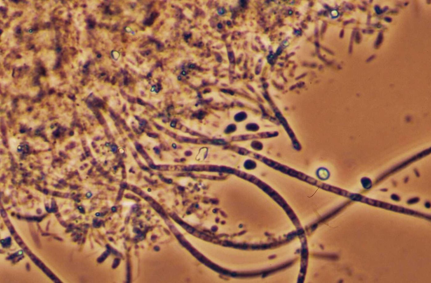
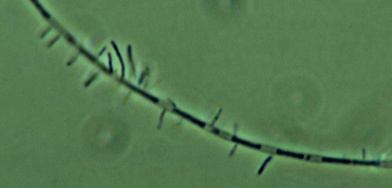
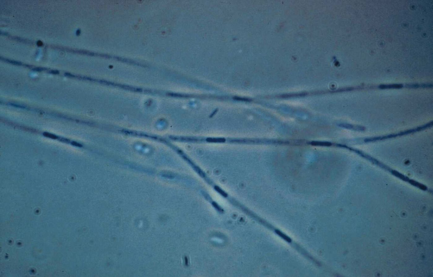
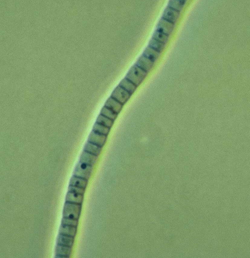
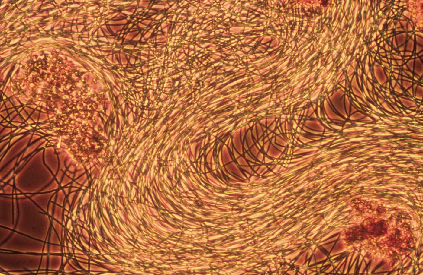
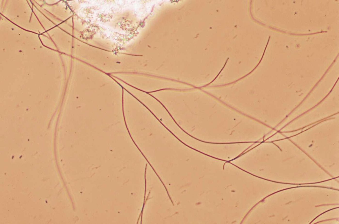
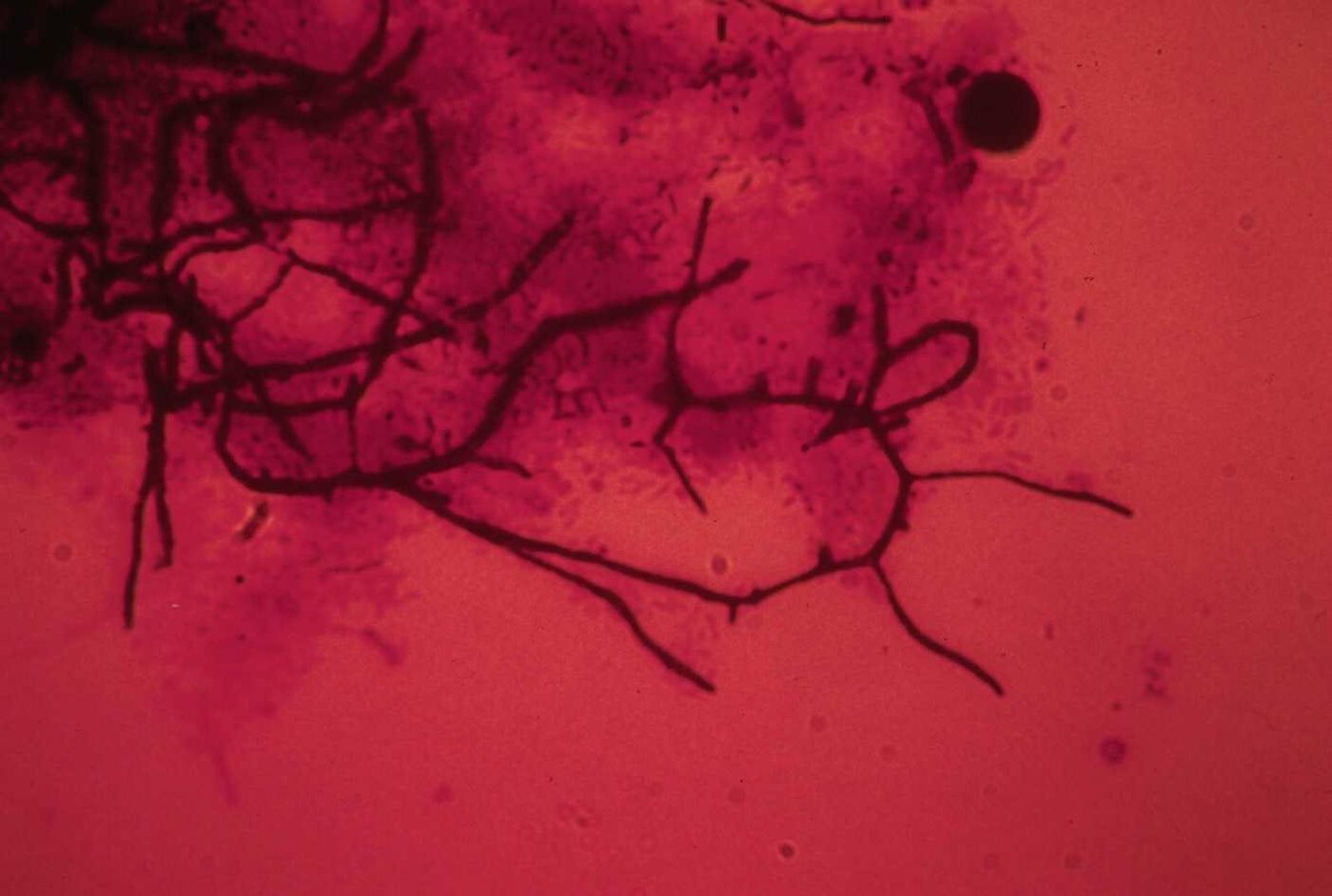
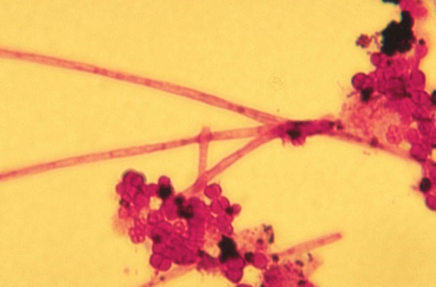
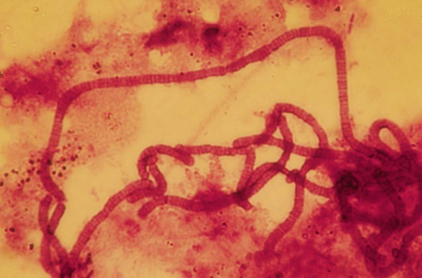
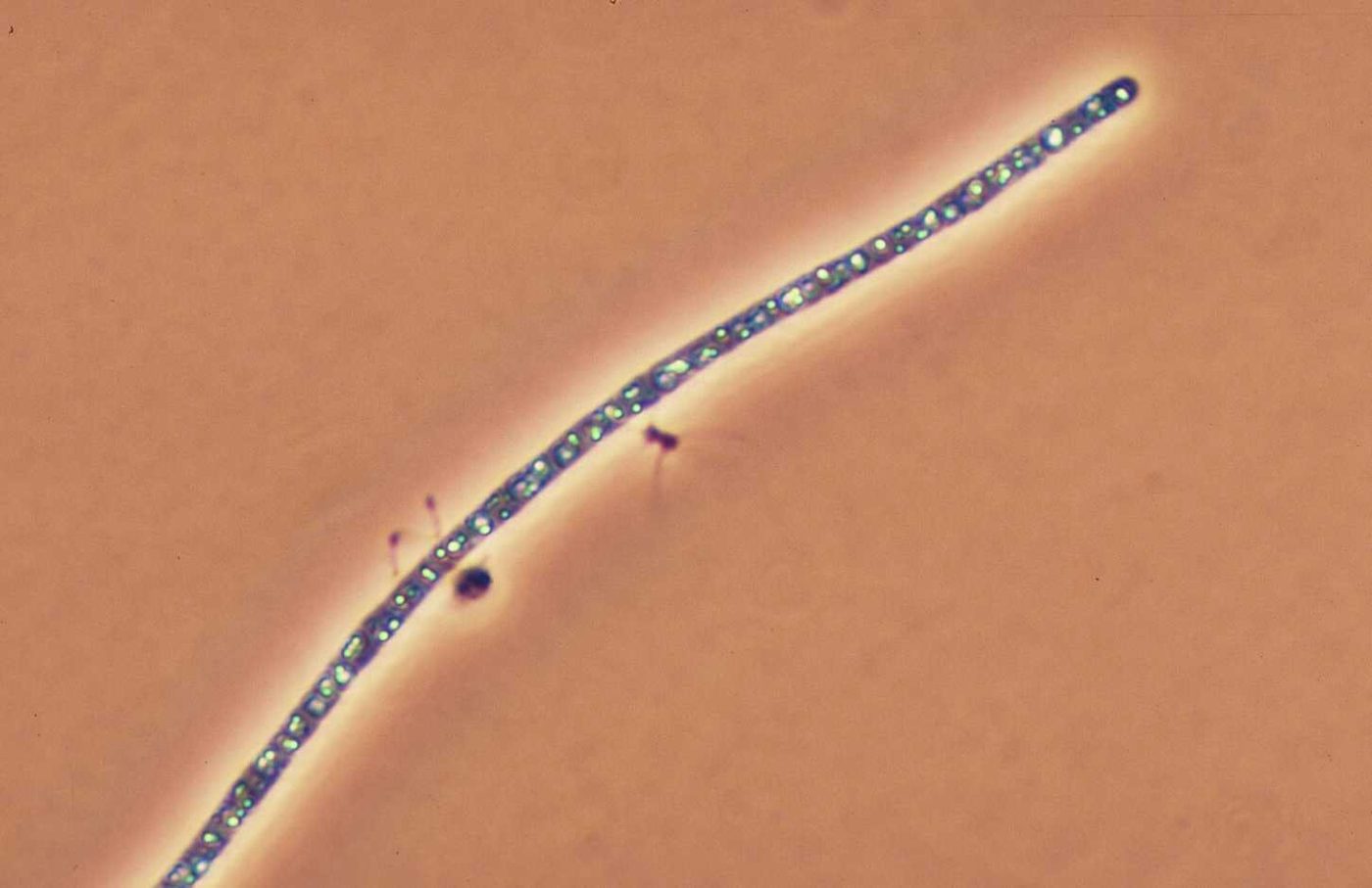
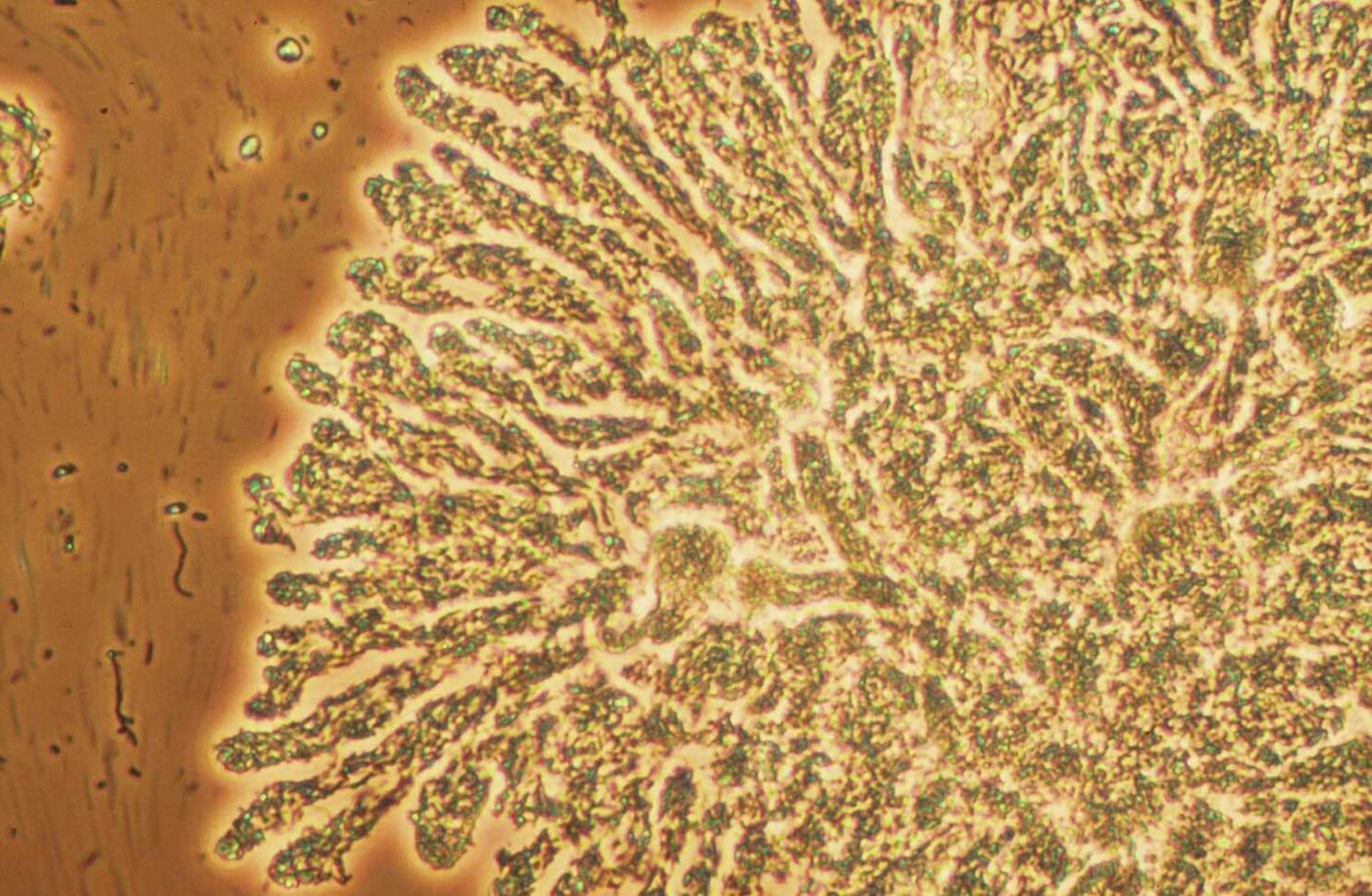
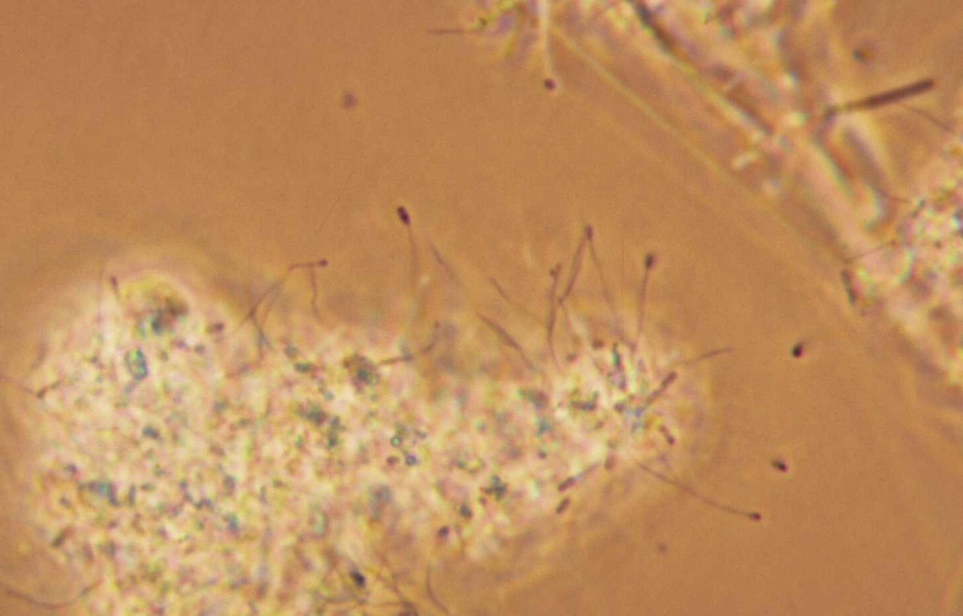
Interfloc bridging. Filaments bridging between flocs. 100x. What this indicates: Filaments bridging floc to floc hold the flocs apart and cause bulking: high SVI and a slow settling blanket.Type 021N. Type 021N in large numbers. Phase contrast, 200x. What this indicates: Type 021N points to nutrient deficiency or septic, high organic acid wastes.Sphaerotilus natans. Sphaerotilus natans, cells inside a clear sheath. Phase contrast, 1000x. What this indicates: S. natans is a classic low DO filament.Nocardioforms, Gram stain. Nocardioforms: Gram positive with true branching. 1000x. What this indicates: Nocardioforms go with grease, long SRT and thick brown foam on the aeration basin.Thiothrix. Thiothrix filaments radiating from a floc. Phase contrast, 1000x. What this indicates: Thiothrix points to septic wastes and sulfide, or to nutrient deficiency.Beggiatoa. Beggiatoa, a sulfur oxidizing filament that glides. Phase contrast, 400x. What this indicates: Beggiatoa indicates septicity and sulfide in the wastewater.S. natans, Type 021N and Type 0041. S. natans, Type 021N and Type 0041 in one field. 1000x. What this indicates: S. natans points to low DO. Type 021N points to septic wastes or nutrient deficiency, and Type 0041 to low F/M or nutrient deficiency.Thiothrix II with chlorine damage. Thiothrix II after RAS chlorination. 1000x. What this indicates: Broken, empty filaments after RAS chlorination mean the dose is reaching them. Watch for floc damage too.Filament with chlorine damage. Filament damaged by chlorine. 400x. What this indicates: Chlorine damage confirms the dose is working on filaments. Check that floc formers are not damaged as well.Damaged S. natans. Damaged S. natans filaments with empty sections. What this indicates: Empty sections in the sheath are a sign of chlorination or another stress.Type 021N, 1000x. Type 021N close up. Phase contrast, 1000x. What this indicates: Nutrient deficiency or septic wastes.Type 021N overgrowth. Type 021N overgrowth caused by high organic acids. What this indicates: Septic, high organic acid wastes can drive 021N to bulking levels.S. natans, 200x. S. natans with false branching. 200x. What this indicates: Low DO filament.Nocardioforms, Gram stain. Nocardioforms, Gram positive and branched. What this indicates: Grease, long SRT and brown foam.Type 0041, Gram stain. Type 0041 with attached growth, Gram stain. 1000x. It stains Gram negative here; Type 0041 is usually listed as Gram positive or variable, and attached growth can hide the reaction. What this indicates: Low F/M or nutrient deficiency.Nostocoida limicola II. Nostocoida limicola II, a filament usually listed as Neisser positive. 1000x. What this indicates: Low F/M, low DO or industrial organic wastes.Thiothrix I. Thiothrix I. Phase contrast. What this indicates: Septic wastes, sulfide or nutrient deficiency.
Illustrated
Capsules and sheaths. Bacteria with slime capsules (clear halos in India ink) and a sheathed filament with cells inside, an empty section and a false branch. What this indicates: Heavy capsule (slime) formation can cause viscous bulking, often from nutrient deficiency; a sheath and false branching help identify filaments such as S. natans.Original illustration for this site, not to scale.
Floc and dispersed growth
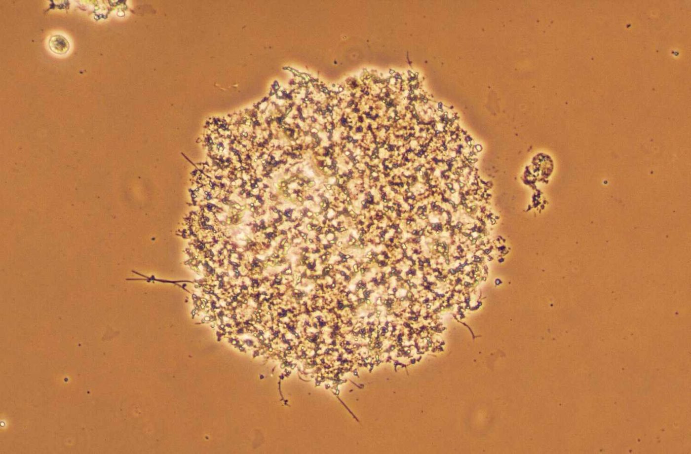
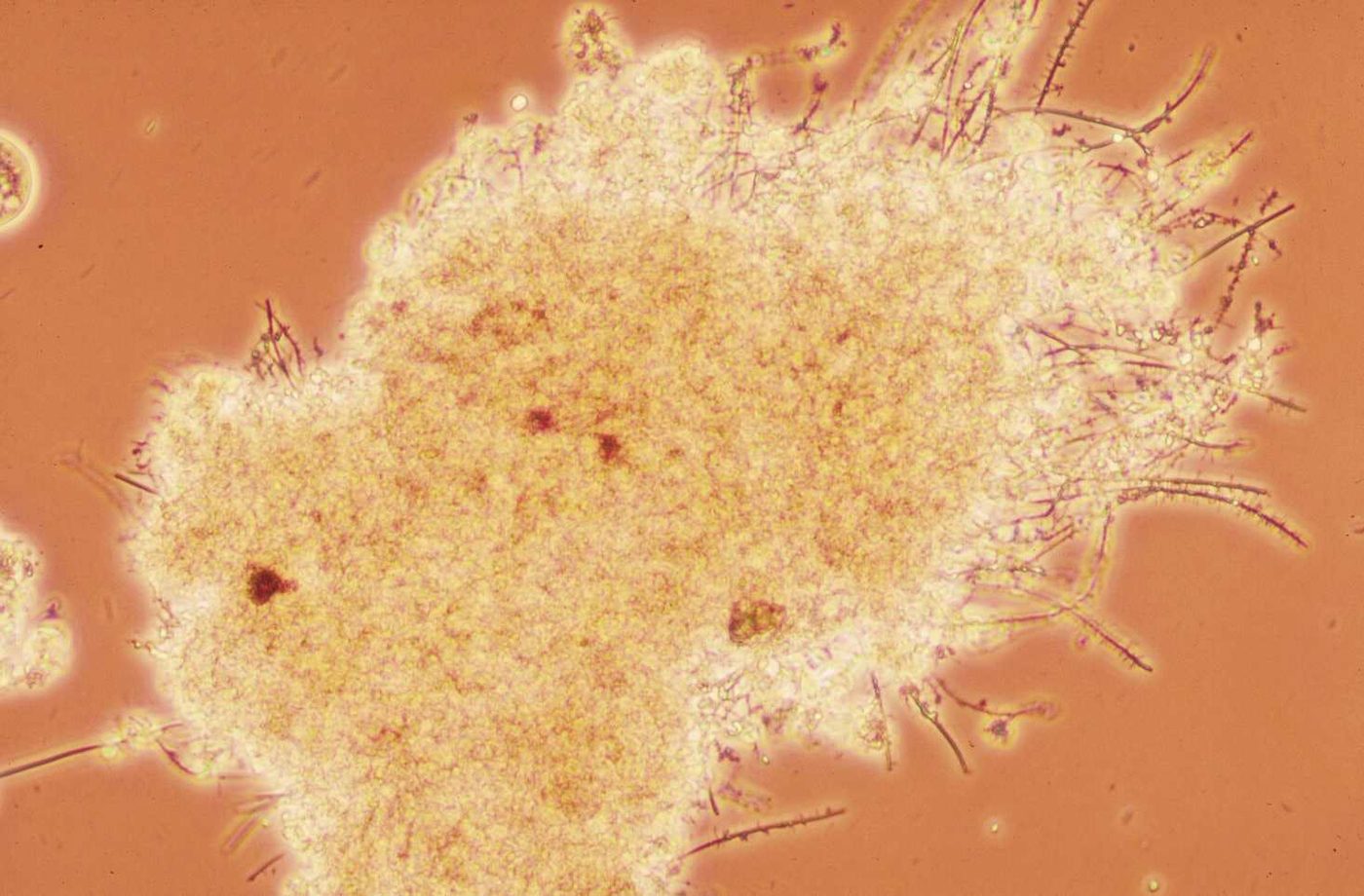
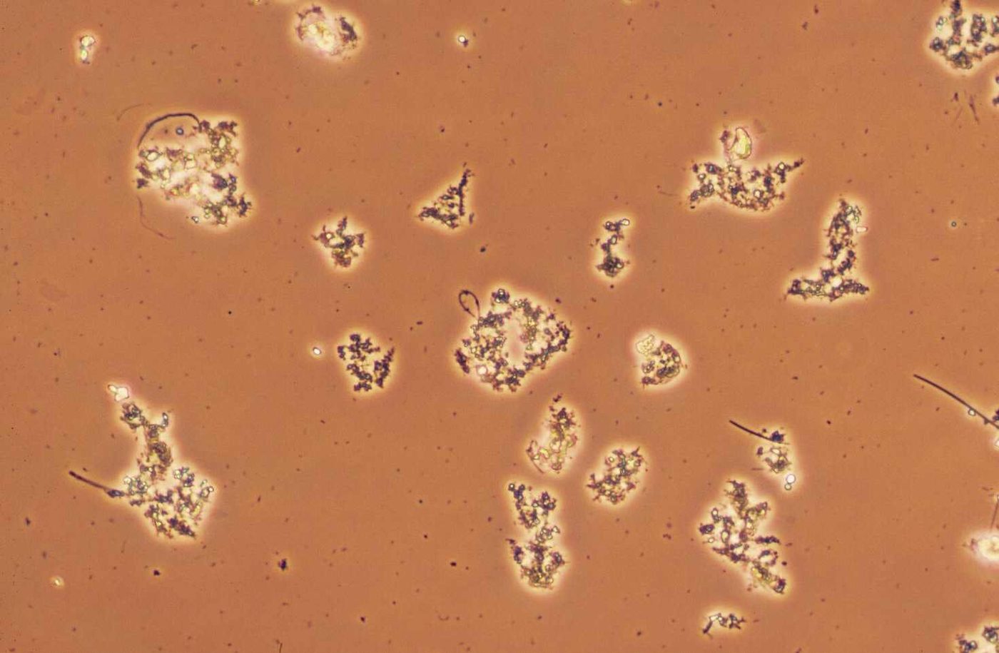
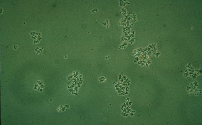
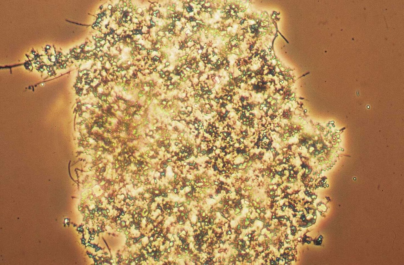
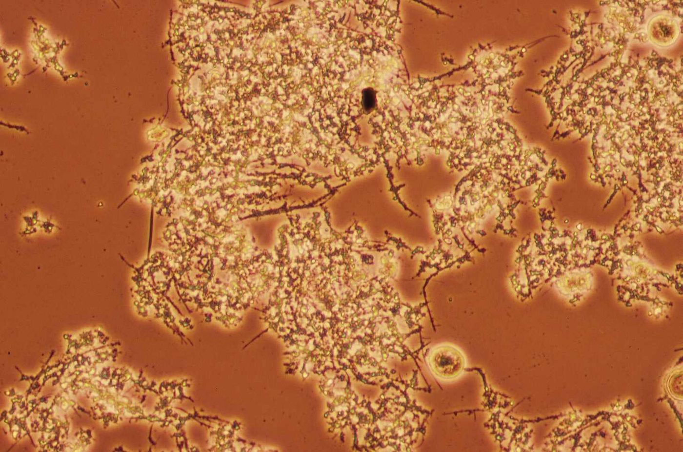
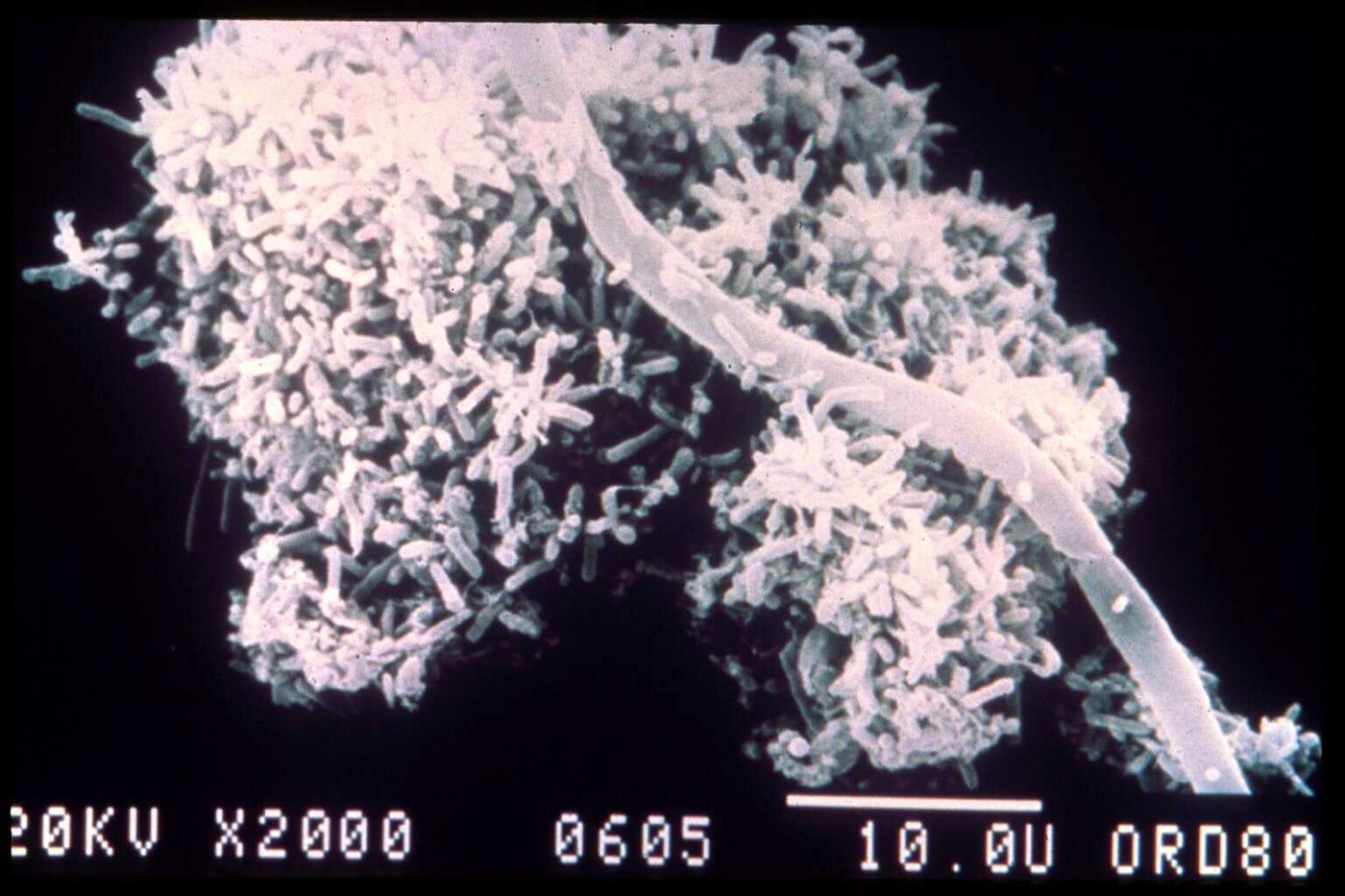
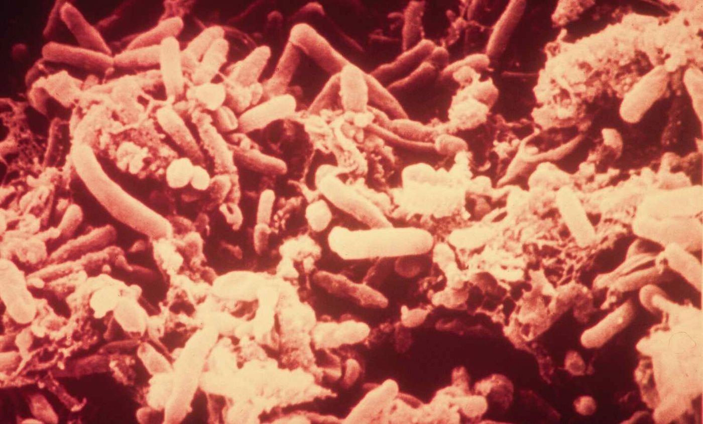
Compact floc. Round, firm floc with few extending filaments and a clear background. Phase contrast. What this indicates: Firm floc with a clear background settles well. If all the floc is this small and round, watch for pin floc.Floc with extending filaments. Large floc with filaments growing out past its edges. Phase contrast. What this indicates: Filaments extending from floc are an early bulking warning. Rate their abundance and track SVI.Small, irregular floc. Small, irregular flocs with a clear background. Low power, phase contrast. What this indicates: Small floc settles slowly and may leave a hazy supernatant. Compare with the settleometer.Pin floc. Pin floc: small, compact particles scattered across the slide. What this indicates: Pin floc occurs at long sludge ages. SVI is often low but the supernatant is turbid.India ink stain. India ink reverse stain of a floc. 100x. What this indicates: Ink fills the background. Large clear areas inside the floc show excess exocellular slime, a sign to check for viscous bulking and nutrient deficiency.Fingered Zoogloea. Fingered Zoogloea. 400x. What this indicates: Found in high F/M systems with soluble, easily degraded food and low pH. It can cause viscous bulking.Open floc. Large open floc with some filaments. Phase contrast. What this indicates: Open floc settles slower than firm floc. Check DO and SRT.Floc without India ink. Floc without India ink, for comparison. What this indicates: Compare with the India ink slide to judge slime content.India ink, several flocs. Several flocs in India ink. What this indicates: Use India ink to check for excess exocellular slime.Dispersed bacteria. Single, dispersed bacteria with almost no floc. What this indicates: This will not settle. Dispersed growth means young sludge, a high soluble load or toxicity breaking floc apart.Straggler floc in the clarifier. Straggler floc carrying over the clarifier weirs. What this indicates: Straggler floc can be hard to tell from pin floc under the microscope. The settling test shows poor settling and a ragged interface.Floc formers and paper fiber, SEM. Floc formers on a paper fiber. Scanning electron microscope, 2000x. What this indicates: Fibers act as a backbone for floc, much like filaments do.Floc formers, SEM. Floc forming bacteria. Scanning electron microscope, 7200x. What this indicates: These rods are what build healthy floc.
Illustrated
Inside a healthy floc. Floc forming rods held in sticky EPS around a short filament backbone, with trapped particles and stalked ciliates on the edge. What this indicates: Firm, irregular floc with a few internal filaments and a clear background settles well and leaves a clear supernatant.Original illustration for this site, not to scale.Healthy floc, bulking and pin floc. Three 100x views, each with its 30 minute settleometer result. What this indicates: Filaments bridging flocs mean high SVI and slow settling; tiny, filament free pin floc settles fast but leaves a cloudy supernatant, often from too long an SRT.Original illustration for this site, not to scale.Dispersed growth compared with healthy floc. A healthy floc field next to a field full of single cells and tiny clumps, with their settleometers. What this indicates: Dispersed growth points to a very young sludge, toxicity, high load or low DO. Expect turbid effluent until floc reforms.Original illustration for this site, not to scale.
Other bacteria
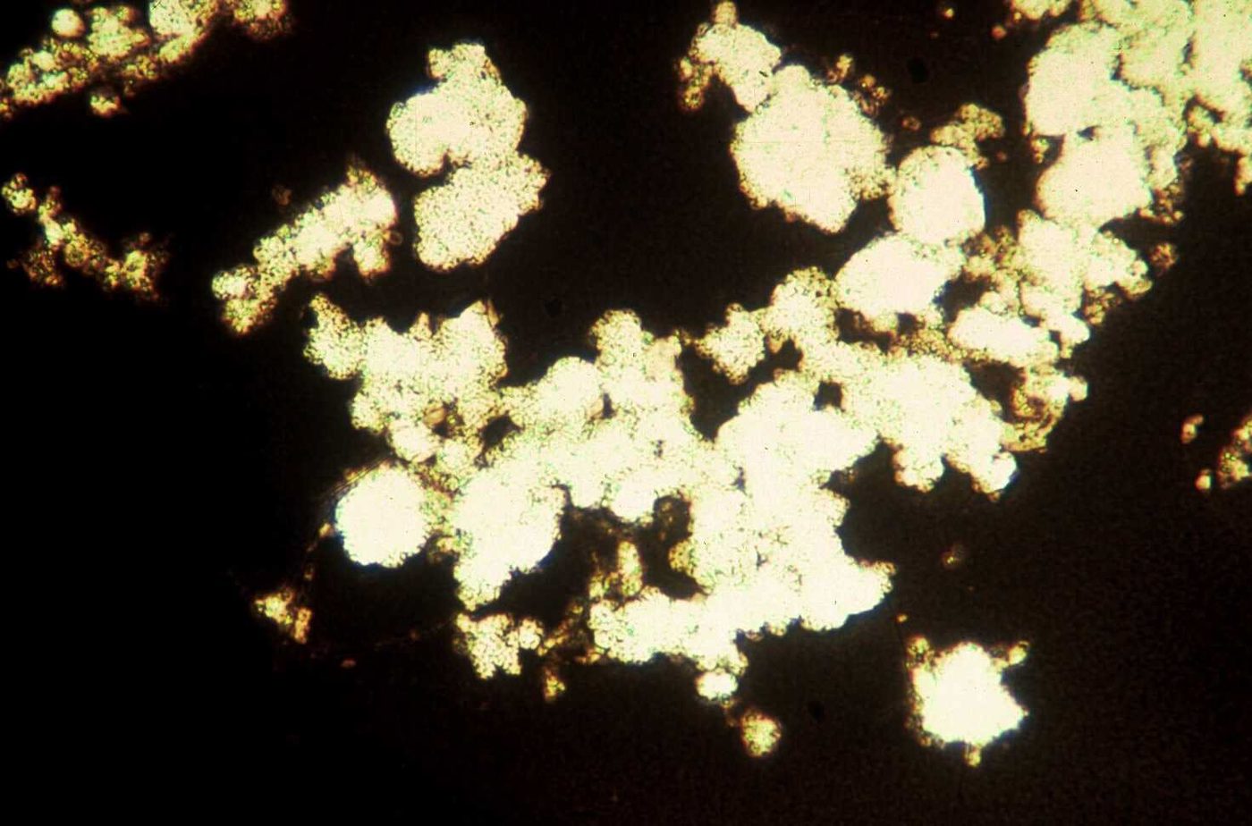
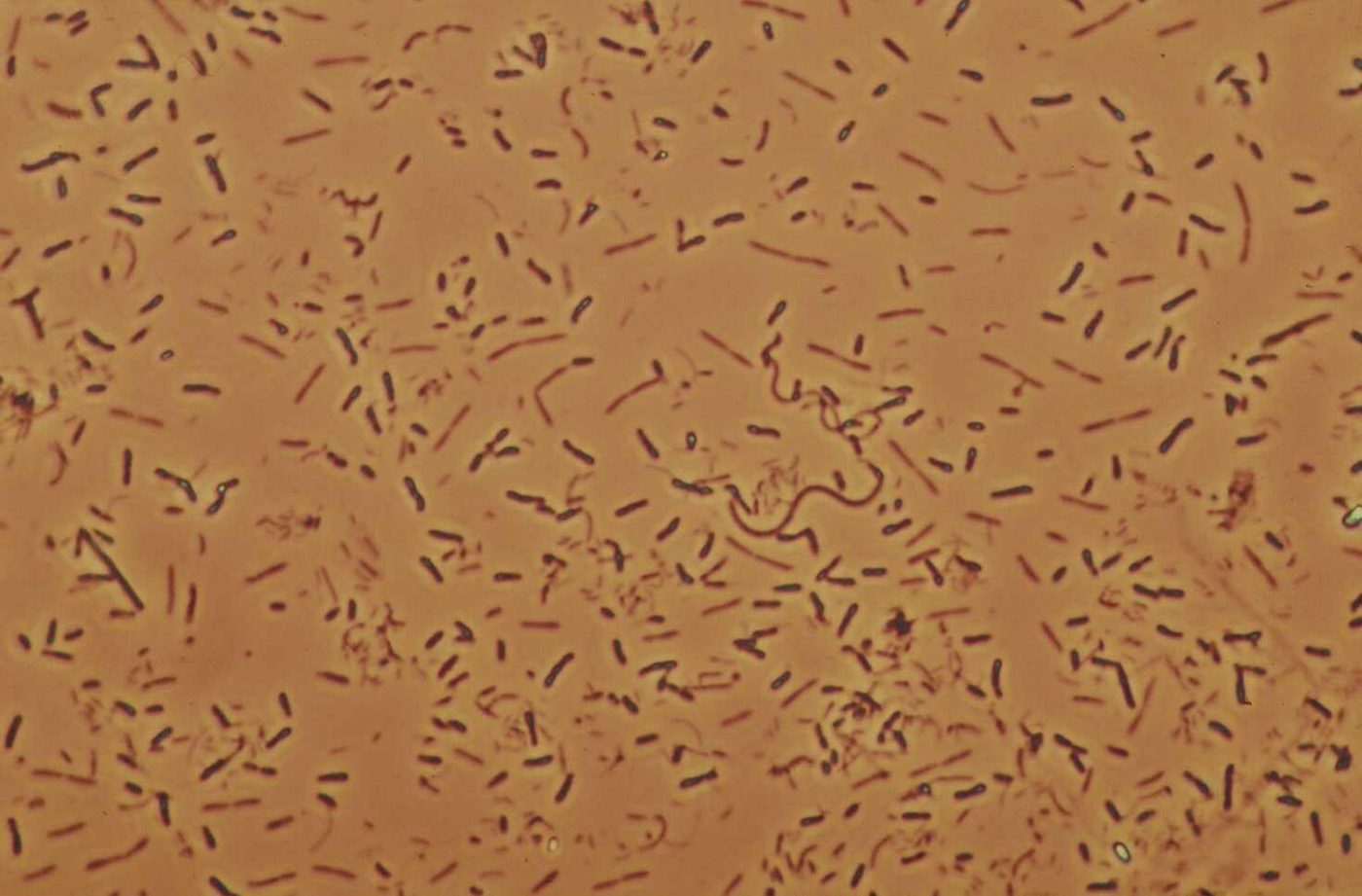
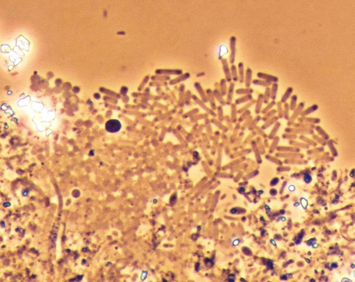
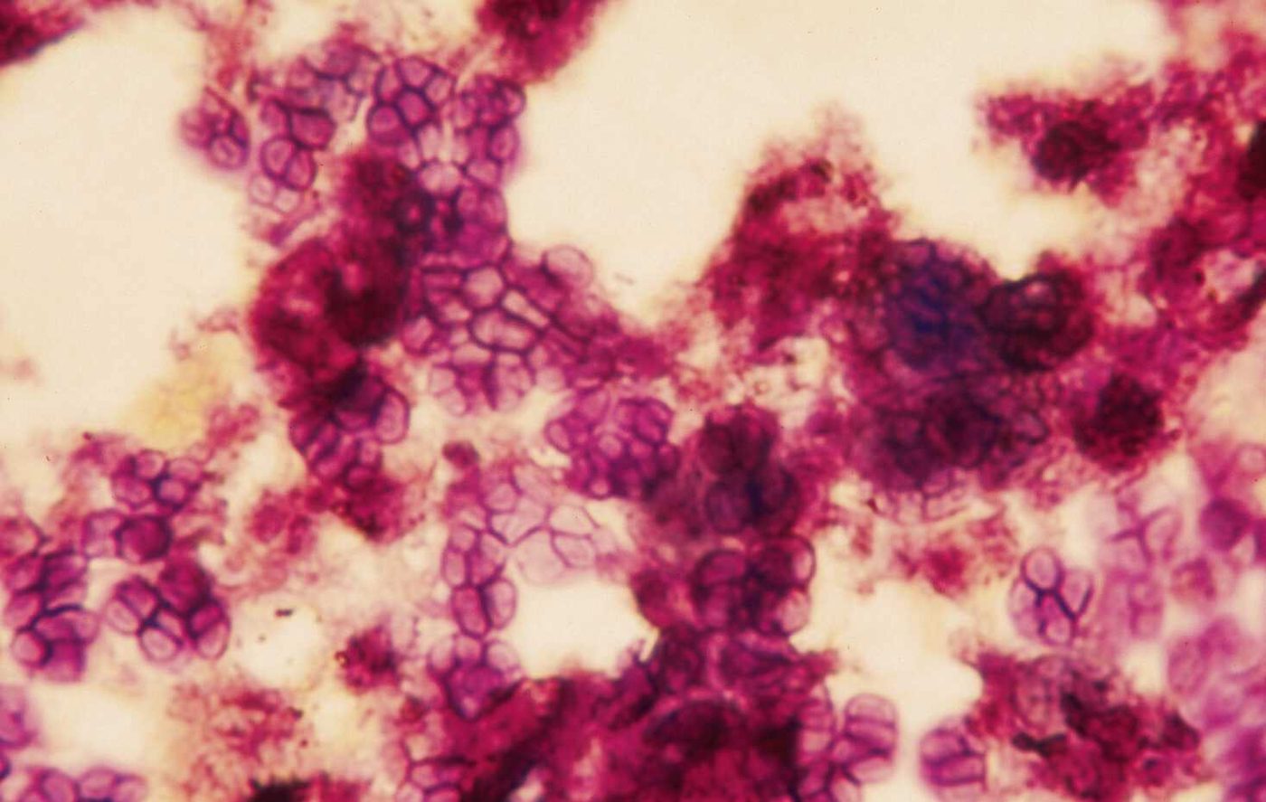
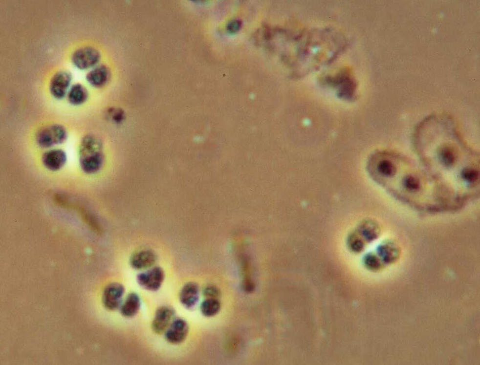
Nitrifying bacteria (as labeled on the slide). Bacterial cluster shown as nitrifying bacteria. Nitrifiers cannot be confirmed by shape alone; that takes a molecular test such as FISH. Phase contrast. What this indicates: Nitrifiers grow slowly and need adequate SRT, DO and alkalinity. In most plants Nitrospira, not Nitrobacter, is the main nitrite oxidizer.Acinetobacter (Bio-P), Gram stain. Acinetobacter type cells, Gram negative stain. What this indicates: Long linked with biological phosphorus removal, though other bacteria (such as Accumulibacter) are now known to do most of it. Often seen with high organic acids.Hyphomicrobium. Hyphomicrobium. What this indicates: Associated with denitrification, especially with methanol addition.Tetrads. Tetrads (cells in groups of four). What this indicates: Tetrads often go with high organic acids in the feed.
Illustrated
Bacterial shapes. Cocci (singles, pairs, chains, clusters, tetrads), bacilli (rods) and spirals at 1000x. What this indicates: Shape alone does not identify a bacterium, but it is the first step in reading a Gram stain, and tetrads in large numbers can point to high organic acids.Original illustration for this site, not to scale.Aerobic respiration. Bacteria use food (organics) and oxygen to make carbon dioxide, water, new cells and energy. What this indicates: This is why oxygen demand rises with load, and why the new cells must be wasted to hold a steady sludge age.Original illustration for this site, not to scale.
One slide is a snapshot. Tie what you see to SRT, F/M, DO, SVI and effluent quality before you change anything. Start with Identify what I'm seeing or the microscopy guide.